PA WRIST PROJECTION
Standard Posteroanterior view for general evaluation of the carpal bones and distal forearm
Radiografía PA Muñeca
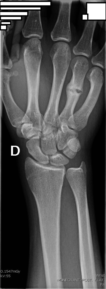
Diagrama de Posición PA Muñeca
Exposure Factors
45-55
Kilovoltage (kV)
2-4
mAs
100 cm
Distance (SID)
Fine
Focal Spot
No
Grid (Bucky)
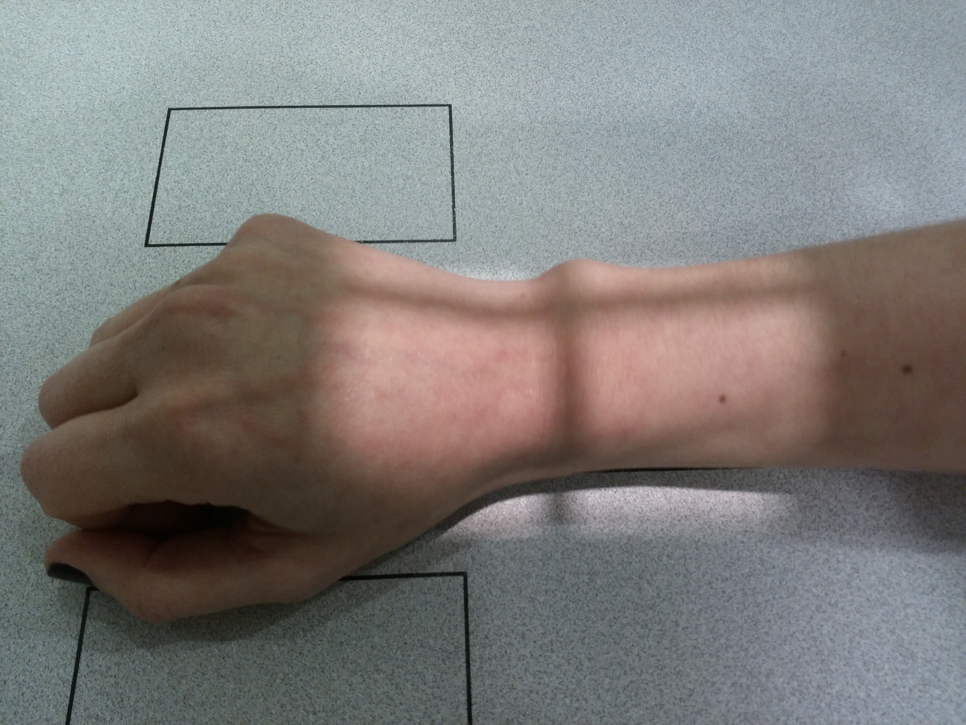
Patient Positioning
Place the hand and wrist in a prone position on the image receptor.
Arch the hand (cup position) to bring the carpus into direct contact with the receptor.
Ensure the distal forearm (radius and ulna) is parallel to the long axis of the IR.
Instruct the patient to remain still during exposure.
Central Ray
Mid-Carpal Area
Location: Perpendicular to the mid-carpal area (midpoint between styloid processes).
Visible Anatomy
Carpal Bones
Visualization of all 8 carpal bones.
Distal Forearm
Distal radius and ulna included.
Metacarpals
Proximal portion of the metacarpals.
ROUTINE PROTOCOL (Split Plate)
PA Projection
On the first half of the plate
• Bone structure evaluation
• Joint space assessment
• Carpal alignment
Lateral Projection
On the second half of the plate
• Displacement evaluation
• Carpal-metacarpal alignment
• Profile visualization
A split plate allows for both necessary projections for a complete wrist evaluation.